ALS, SMA and other motor neurone diseases
Special consultation ALS, SMA and other motor neurone diseases
In the special consultation hour for motor neurone diseases, we deal with clinical and scientific issues of motor neurone diseases.
- Nationwide highly specialized patient care (incl. NIV, IV ventilation, artificial nutrition, eye control computer)
- Involvement in national and international cohorts
- drug studies
- Studies with clinical questions
- basic scientific studies
In this special consultation, outpatients with motor neuron diseases, in particular amyotrophic lateral sclerosis (ALS), are diagnosed and treated. Medical treatment is supplemented by socio-medical care and related medical subjects, especially pulmonary medicine (mechanical respiratory aids), swallowing diagnostics and neurogenetics. If necessary, inpatient stays are organized for diagnostics and therapy.
Consultion time:
By appointment
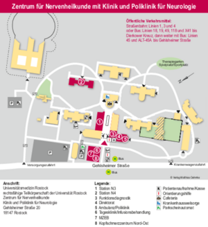
Neurology Polyclinic
Gehlsheimerstrasse 20
18147 Rostock
Link to the map of the outpatient clinic
Appointment arrangement:
(0381) 494 5276
(0381) 494 9798
Case management Mecklenburg-Vorpommern:
Sophie Fischer
Neurology Polyiclinic
Sophie.Fischer{bei}med.uni-rostock.de
(0381) 494 149542
- Ensuring outpatient care
- interdisciplinary cooperation with medical and medical aid providers, health insurance companies, patient organizations etc.
Your contact person

Prof. Dr. Dr. Andreas Hermann
Section for Translationale Neurodegeneration "Albrecht Kossel"

Prof. Dr. med. Johannes L. Prudlo
Experimental Neurology, research area ALS
![[Translate to English:] -](/fileadmin/_processed_/9/3/csm_Bild_Kamm__Christoph_2d6435f9ee.jpg)
PD Dr. med. Christoph Kamm
Experimental Neurology, research area ALS
![[Translate to English:] -](/fileadmin/_processed_/6/c/csm_sophie_6_78d6c06864.jpg)
Sophie Fischer
Case management Mecklenburg-Vorpommern
Studycoordinator & Study Nurse
sophie.fischer@med.uni-rostock.de
Amyotrophic lateral sclerosis (abbreviation: ALS) is one of the so-called motor neuron diseases and is a degenerative disease of the motor nervous system, i.e. the nervous system that is responsible for all voluntary movements. It is also called amyotrophic lateral sclerosis or myatrophic lateral sclerosis. It is often referred to in English as Lou Gehrig's syndrome, after a 20th-century baseball player, or Charcot's disease after its first describer, Jean-Martin Charcot.
Epidemiology
ALS has an incidence of 1.5-2.6/100,000 and a prevalence of 6-8/100,000 population. Males are more commonly affected than females, with a ratio of 1.5-2:1. Most cases occur between the ages of 40 and 70 (mean age 58 years), with the earliest cases observed at the age of 20. The incidence increases with age. The course is rapidly progressive. Approximately 10-15% of patients survive 10 years, isolated cases of patients surviving >40 years have been described. The bulbar form usually has a worse prognosis (mean survival 2-2.5 years). However, there are large inter-individual fluctuations that do NOT allow a reliable prognosis for the respective patient.
Physiology
In ALS there is a combined degeneration of the 1st and 2nd motor neurons. The muscles that are responsible for voluntary movement (so-called striated muscles) are innervated by so-called motor neurons. Each muscle is supplied by its own second motor neuron (α-motor neuron), the cell body of which is located in the spinal cord and only the extension of it extends to the periphery. The longest extensions are those that extend from the spinal cord of the lumbar spine to the muscles of the foot, which are >1 m in length. The cell bodies of the second motoneurons are distributed over the entire length of the spinal cord, depending on which muscles are innervated by them. For example, the motoneurons for the arm muscles are in the area of the lower cervical cord, those for the torso muscles in the area of the upper thoracic spine and those for the leg muscles in the area of the lumbar cord. Every 2nd motor neuron is innervated by a 1st motor neuron (so-called Betz pyramidal cells), which is located in the motor cortex in the cerebrum. From these, in turn, they only send out their extensions in the direction of the spinal cord. Each 1st motor neuron is connected to a special, separate 2nd motor neuron. Smooth functioning of the respective 1st and 2nd motor neurons is essential for every movement.
Pathology
If the second motor neuron degenerates, the patient notices a flaccid paralysis of the corresponding muscle. In addition, so-called fasciculations, muscle twitching, can occur. In the course of this, there is a significant decrease in muscle mass (atrophy). A degeneration of the 1st motor neuron alone often does not result in paralysis in the actual sense, but in increasing stiffness of the muscles (so-called spasticity). Patients often first notice this when climbing stairs or when walking unsteadily, up to severe limitations and pain with every movement. In addition, the muscle reflexes become very lively. ALS is a combined degeneration in which there are large interindividual differences in the involvement of the 1st and 2nd motor neurons.
Disease/course
Clinically, a distal, unilateral extremity atrophy is the most common initially. It is only later that other muscles are affected and the picture is paralyzed on both sides, later affecting all extremities. About 1/3 of the patients have a so-called bulbar form. This involves paralysis of the swallowing and speech muscles, sometimes also associated with a weakness in holding the head. Only as the disease progresses does increasing paralysis of the arms, legs and, above all, the respiratory muscles occur. In both forms of the disease, the eye muscles and the muscles of the anus are usually spared. It is still not known why these are not affected. The final stage of the disease is characterized by the attack on the respiratory muscles. Patients usually die from respiratory failure. Often there is also pneumonia caused by swallowed saliva or food (aspiration pneumonia). The bulbar form is therefore the one with the poorer prognosis because the respiratory muscles are often affected early on without paralysis of the extremities necessarily occurring. As the disease progresses, sooner or later all voluntary muscles are affected, i.e. not only paralysis of all extremities but also speech and swallowing disorders up to the inability to communicate and eat. What all forms have in common is that patients are usually fully conscious to the end. About 20% form a dementia syndrome of varying degrees.
Special forms
If only or primarily the 1st motor neuron is affected, this is referred to as primary lateral sclerosis. The prognosis for this is significantly better. In some cases, both arms become paralyzed with no signs of further paralysis. This is known as the so-called "flail arm syndrome". Both forms described have in common the transition to ALS in full development over the course of the disease. If only the 2nd motor neuron is affected, this is referred to as spinal muscular atrophy. These are usually hereditary and in most cases occur in childhood.
Reasons
The cause of the selective degeneration of only the motoneurons is not known to this day. There are numerous theories (not detailed here) that involve mitochondrial, cytoskeletal, oxidative stress, inflammatory responses, or neurotransmitter imbalances. What they all have in common is that no clear causal connection between all ALS diseases has been established for any theory to date. The main problem in research to date is that it is so difficult to examine the diseased cells because they cannot be taken from the human without causing serious damage. New techniques, such as the so-called induced pluripotent stem cells, could possibly make this possible in the future.
ALS is a sporadic disease. Only about 5-10% of cases are hereditary. One also speaks of familial ALS syndromes. Of these 5-10%, around 20% have mutations in the SOD1 gene (copper-zinc-superoxide-dismutase). For a long time, this was the only known gene that causes ALS. Therefore, until recently, the animal models on which most drugs were tested were based on mutations in the SOD1 gene. A lot has happened in the last five years. Three other genes were discovered, angiogenin (ANG), TDP-43 (synonym TARDP) and FUS. ANG is responsible for about 2%, TDP-43 for 1-4%, FUS about 5% of familial ALS diseases. Interestingly, 1-5% of patients without a familial cluster also show mutations in the TDP-43 or FUS gene. In summary, the genes responsible for >50% of familial ALS diseases remain unknown.
Data from pathological examinations recently revealed an interesting piece of news. The spinal cords of patients with sporadic ALS were compared with those of patients with gene defects in SOD1, FUS and TDP-43. It turned out that the pathological changes of all forms were similar except for those suffering from SOD1 mutations. This could mean that until recently the only available animal models of the disease (those with SOD1 mutations) may not reflect the disease process/etiology of most ALS diseases. It will be interesting to see whether newer animal models will be better for testing new drugs.
Even today, the diagnosis of ALS is a diagnosis of exclusion. A final definitive diagnosis can strictly speaking only be made by a pathologist post mortem. Even today, there are still no markers or detection methods that allow ALS to be clearly diagnosed at an early stage.
As a standard procedure, imaging of the head and cervical spine using magnetic resonance imaging should be performed when progressive and spreading paralyses occur. However, in addition to the clinical examination, the most important examination is the electrophysiological measurements. This involves measuring both the nerves (electroneurography, ENG) and the muscles (electromyography, EMG). In ENG, nerve damage should be excluded as the cause of the paralysis (e.g. polyneuropathies, herniated discs, post-polio syndrome). It is particularly important to search for so-called conduction blocks, which would be typical of an autoimmune-mediated nerve disease (multifocal motor neuropathy, MMN). Using needle recordings of the musculature (EMG), a distinction can be made between muscular and neuronal causes. The involvement of the 1st motor neurone is measured using motor-evoked potentials.
Inflammatory processes should be ruled out (e.g. Lues, Lyme disease and, in rare cases and with a corresponding medical history and clinical findings, Creutzfeld-Jakob and AIDS-mediated nerve diseases). If there is a corresponding history, a tumour search should be carried out to rule out paraneoplastic syndromes (e.g. a breast tumour should be ruled out, particularly in women with a predominant involvement of the 1st motor neurone).
For further details on diagnostics, please refer to the recommendations of the German Neurological Society:
To date, there is no cure for ALS. Only one drug has been approved for the treatment of ALS, riluzole. This inhibits the effect of the neurotransmitter glutamate, which has been shown in studies to delay the progression of the disease. A critical point to note is that not every patient tolerates this medication well and the delay in disease progression was only a few months on average. However, the effect for each individual patient cannot be predicted and therefore the therapy should not be withheld from any patient.
Numerous drugs have been tried. What they all had in common was an effect in animal models that could not be confirmed in humans. One reason for this could be that the animal models used to date (mutation in SOD1 gene) did not represent the true course and cause of the disease. New animal models and alternative technologies (e.g. induced pluripotent stem cells) to produce more reliable model systems are urgently needed. Our working group is particularly concerned with new cell systems and, in cooperation with the working group of Prof. Dr Albert Ludolph from Ulm, with new animal models.
The major dilemma of ALS treatment is the following: as ALS is still a diagnosis of exclusion (see Diagnostics), an early diagnosis can almost never be made with absolute certainty, but only during the course of the disease, i.e. when some muscles are already paralysed. As a doctor, you only want to make such a fatal diagnosis when you are as certain as possible. This makes it difficult to start treatment at an early stage.
This applies, among other things, to treatment with riluzole. As the so-called glutamate-mediated cytotoxicity that riluzole treats first leads to damage and then to the destruction of the respective motor neurone, early treatment - as long as the nerve cells are only damaged - is obligatory. Nerve cells that have already perished can no longer be saved.
To this day, the mainstay of treatment is adjunctive or symptomatic therapy, i.e. treatment of symptoms/accompanying symptoms and prophylaxis of complications.
Physiotherapy
Every patient should receive regular physiotherapy. There are no fixed rules; each patient must develop their own therapy plan with their physiotherapist. Each patient must also work out the frequency of treatments per week for themselves. As a rule, this is 1-2x/week. Physiotherapy has many different tasks. In addition to maintaining the greatest possible mobility, it should in particular accompany and adapt to the disabilities that arise in the course of the disease and the necessary aids. Physiotherapy is also essential at an advanced stage in order to avoid contractures caused by immobility.
A frequent question from patients is how much strain they can put on themselves. It is important to know that ALS is not like a memory that is getting emptier and emptier and should therefore be spared. On the contrary, you should try to counteract muscle loss for as long as possible by exercising regularly. However, this does not mean always exercising to the limit and beyond. A good balance is the right solution here.
Logotherapy
Logotherapy becomes more important as soon as the speech and swallowing muscles become involved. Every patient who notices the first symptoms in this regard should start with it and also endeavour to receive regular care in this area. This includes improving articulation and swallowing, providing aids or tricks to change food intake and supporting patients with communication aids (e.g. communicator, eye control computer). Our experience here has been consistently good. Here, too, the principle applies that the patient must see and learn for themselves to what extent they benefit from the respective therapy.
Ergotherapy
Occupational therapy is particularly important for maintaining fine motor skills and in the course of providing aids. For example, maintaining writing and hand activities is an important domain of occupational therapy. Here too, the individual patient decides whether therapy is necessary.
Supply of medical aids
The provision of aids is a key factor in the course of the disease. It is important that patients and relatives act with foresight. As medical aids are often expensive, they must first be applied for from the health insurance company. However, such an authorisation procedure can be very lengthy. It is not uncommon for us to receive authorisation when the aid was no longer needed because the illness had already progressed significantly. Further details on the provision of aids can be found here.
Muscle cramps (Crampi)
Cramps in individual muscles (e.g. calf cramps) are often a non-specific early symptom of ALS. The cramps can also affect several muscles at the same time. They sometimes occur spontaneously, sometimes after exertion. In addition to regular physical training or physiotherapy exercises, drug therapies, e.g. with magnesium, quinine sulphate or carbamazepine, can help.
Dysphagia
The weakness of the throat and pharyngeal muscles leads to swallowing disorders in ALS patients (so-called dysphagia). The main task of the normal swallowing process is to prevent parts of the ingested food or liquids (saliva) from entering the respiratory tract. Disorders result in frequent swallowing or "not getting food down". Pneumonia (so-called aspiration pneumonia) often develops as a result of liquids entering the lungs. If these symptoms occur, it is often necessary to change the diet, e.g. to pureed food and thickened liquids, in addition to treatment with speech therapy. High-calorie food supplements can be used to cover the body's energy requirements. Artificial feeding via a so-called PEG tube is often necessary during the course of the disease. This should be inserted at an early stage, as the rate of complications during insertion is higher in patients with advanced respiratory muscle weakness, including a significantly increased risk of the necessary anaesthesia. The advantage of the horizontal PEG tube is that oral feeding is still possible even after a feeding tube has been inserted, but the pressure to consume sufficient calories is reduced. However, inadequate calorie intake should be avoided at all stages of the disease.
Salivation
Another consequence of dysphagia is increased saliva loss from the mouth (sialorrhoea). Strictly speaking, this is not normally an increased flow of saliva but a swallowing disorder of normal amounts of saliva. There are medications that reduce salivation (e.g. amitriptyline or scopolamine). Belladonna tincture also works reliably, but patients with glaucoma should not take this substance. The quarterly injection of botulinum toxin into the salivary glands is also an effective treatment option.
Speech disorders and communication
As the disease progresses, the muscles responsible for articulation usually become increasingly paralysed (dysarthria). In addition, increasing breathlessness can make speaking even more difficult. The restriction of communication with the environment is often one of the most distressing symptoms for patients and relatives in the advanced stage. Early speech therapy (speech therapy) treatment is important. In addition, early provision of communication aids is also recommended. Numerous options are available, from alphabet and picture boards to computerised communication aids. In the case of advanced paralyses, computer systems controlled by eye movements have also been established.
Pathological laughter and crying
Pathological laughter and crying refers to sudden laughter or crying by the patient that is inappropriate to the situation. It is a symptom that is often overlooked but nevertheless not uncommon. It occurs in up to 50 per cent of ALS patients. Drug treatment options for this would be amitriptyline or serotonin reuptake inhibitors.
Respiratory disorder
Advanced ALS regularly leads to weakness of the respiratory muscles. The reduced lung function leads to a reduced oxygen content in the blood (hypoxaemia) and - due to reduced exhalation - to an increased carbon dioxide content in the blood (hypercapnia). Typical symptoms are sleep disorders, morning headaches, daytime tiredness, concentration problems, restlessness or, last but not least, shortness of breath (dyspnoea). Infections of the respiratory tract can further worsen the function of the lungs. Respiratory weakness is the most important cause of limited life expectancy in ALS patients.
If symptoms occur, it is possible to adapt so-called non-invasive ventilation (home ventilation). It is important to distinguish this from invasive ventilation. Non-invasive ventilation is machine-supported ventilation that takes place via a breathing mask. Most patients use this type of ventilation. There is clear data that non-invasive ventilation alleviates the symptoms of lung dysfunction and improves quality of life. Nevertheless, residual function of the patient's own breathing is necessary for this, in particular keeping the throat and pharynx open cannot be completely replaced by non-invasive ventilation. Therefore, in the case of advanced respiratory insufficiency, invasive ventilation via a tracheostoma (after a surgical tracheotomy) is necessary sooner or later.
The option of non-invasive or invasive mechanical ventilation should be discussed as a treatment option early on in the course of the disease. Approximately 20-25% of patients opt for nocturnal non-invasive ventilation, less than 10% for invasive ventilation. The decisive factor for the rejection of invasive ventilation by a large number of patients is the feeling of giving up self-determination over the last hours of life and complete dependence on devices. Here, care by means of eye-control computers and networking of household appliances, together with the recently changed legal situation regarding switching off life-sustaining devices, could lead to a rethink. Nevertheless, this should always be decided by the patient's relatives and with the help of detailed advice from experienced centres. We offer appropriate counselling.
RELYVRIO
On 29 September 2022, the US Food and Drug Administration (FDA) approved AMX0035 (sodium phenylbutyrate and ursodoxicoltaurine) for the treatment of adults with amyotrophic lateral sclerosis (ALS) under the brand name RELYVRIO.
Marketing authorisation was applied for in the European Union (EU) and initially not granted, with the reasoning that the results of the Phase III trial (PHOENIX trial) should be awaited first. These were not available until early 2024. The Phase III trial was negative in all study objectives.
As a result, it was clear that RELYVRIO would not be authorised in the European Union (EU). The company Amylyx also withdrew the marketing authorisation in the countries where it had already been granted (USA, Canada), meaning that this therapy is no longer available and cannot be recommended based on the current study situation.
TOFERSEN (Qalsody®) approved for patients with SOD1-ALS in Europe
TOFERSEN (Qalsody®) is an antisense oligonucleotide that binds the so-called messenger RNA of SOD1 (superoxide dismutase 1) and thus significantly reduces the expression of the SOD1 protein. In a pivotal study on SOD1 patients with different mutations in the SOD1 gene, not only was there a reduction in the axonal damage maker neurofilament, but also a slowing of motor decline, measured on the ALS-FRS scale, as well as a prolongation of survival. The drug had therefore been approved by the FDA for the treatment of SOD1-ALS since 2023. Previously, SOD1 patients in Germany had the opportunity to participate in an open access programme run by Biogen. Around 40 patients across Germany have already been successfully treated in this programme. In May 2024, the EMA has now also granted marketing authorisation for Europe. This means that the open access programme will end in June 2024 and the drug will be available in the classic form from 1 July 2024.
Similar to ASO therapy for spinal muscular atrophy (Spinraza®), the drug must be injected into the spinal canal (intrathecal administration). This is done 3 times in the first month, then at 28-day intervals. Experience to date in the German network shows that the treatment is well tolerated by most patients.
Rasagilin (Phase II)
The neuroprotective potential of rasagiline, a monoamine oxidase B inhibitor, is already known from Parkinson's research and has also been evaluated positively in an ALS mouse model. In order to investigate the effect of a daily dose of rasagiline combined with riluzole, a total of more than 250 patients were recruited for a study. Positive effects of treatment were observed in patients with rapidly progressing ALS; In addition, the drug was well tolerated.
The Results were published in Lancet Neurology in 2018.
Tyrosinkinaseinhibitor Masitinib at sporadic ALS
Masitinib is an orally available protein tyrosine kinase inhibitor that inhibits C-KIT, PDGFR and FGFR3, among others. It also shows slight inhibition of ABL and C-FMS. This tyrosine kinase inhibitor, marketed by AB SCIENCE in France, was recently tested in a phase 2 study in patients with ALS. The results, presented to the public for the first time at the ENCALS meeting in Ljubljana in May 2017, sound promising. In a 48-week study against placebo, the 4.5 mg/kg body weight/day group showed an approximately 3 point lower deterioration in the ALSFRS-R and a significantly lower deterioration in quality of life in the ALSAQ40 questionnaire compared to the placebo group . There was also a significant slowing of the deterioration of vital capacity. So far there have probably been no relevant differences in terms of survival. There were slightly more AE's and SAE's in the treatment group, which focused on laboratory safety parameters, the respiratory tract and neurological complaints. The therapy was carried out as an add-on to the riluzole treatment. Masitinib has received so-called orphan drug status from the FDA and the European Medicines Commission (EMA). A so-called conditional marketing strategy was submitted to the EMA in September 2016. A further confirmation study is currently being planned, which will also begin in Germany at the end of 2017.
Masatinib could therefore represent another promising treatment option for sporadic ALS. However, these data should still be viewed with caution, as they have not yet been published in a scientific journal and therefore have not yet been evaluated according to international standards.
Nusinersen (SMA)
Spinal muscular atrophy is a monogenetic disease with a mutation in the SMN gene. The FDA has approved the drug Spinraza ® (nusinersen) in the USA for the treatment of all forms of SMA. On June 1, 2017, the EMA (European Medicines Agency) granted approval for all forms of 5q-associated spinal muscular atrophy.
This is a gene therapy in which the SMN2 gene is changed so that it functions like an SMN1 gene and thereby significantly increases or almost normalizes the activity of this gene or protein. Clinical studies have shown a clear slowing of disease progression and an increase in survival rates in SMA1 children.
The treatment requires injection of the medication into the spinal canal, which is given every 14 days in a saturation phase. As you progress, the intervals can be extended further.
The Rostock University Medical Center and in particular the Neurology Clinic and Polyclinic have been successfully providing adult SMA patients with the new therapy right from the start and are one of the experienced German-speaking centers for this.
----------------------------------------------------------
DGM Recommendation 23.10.2017:
Treatment indication adults
All patients with a molecular genetically confirmed SMA 5qshould be treated if therapy is requested and after detailed medical information, if there are no medical contraindications. A lumbar puncture must be possible as Spinraza® is only approved for intrathecal use through lumbar puncture.
----------------------------------------------------------
GBA Evaluation 21.12.2017:
After reviewing the data, a “significant added benefit” was found for infants with early-onset SMA (Type I). These patients usually show the first symptoms by the age of six months. For small children, whose first symptoms usually appear before the age of two (type II), the benefit is “considerable” and “not quantifiable” for later onset SMA (types III and IV).
Regardless of the benefit assessment, nusinersen can be prescribed for all patients with 5q-SMA.
In amyotrophic lateral sclerosis (ALS), there is progressive loss of both the 1st and 2nd motor neurons with the main symptom of painless muscle weakness with intact sensitivity. Depending on the course, sooner or later the bulbar muscles become involved and ultimately respiratory arrest occurs due to paralysis of the respiratory muscles. The only one currently Approved drug therapy (riluzole, approved as Rilutek®) delays the progression of the disease, but this drug is only effective to a limited extent. A complete halt to the progression or a cure is not possible. As a result, the search for alternative methods is greater than perhaps for almost any other neurological disease.
A consideration that has recently come into the public eye is stem cell therapy.
First of all, we would like to point out that, in our opinion, there is currently NO sufficient data available to recommend stem cell therapy for ALS patients and we also refer to statements from the Deutschen Gesellschaft für Neurologie bzw. des Kompetenznetzwerkes Stammzellen Nordrhein-Westfalens (siehe auch F.A.Z. Sonntagszeitung, 11.10.2010, Nr. 43, Seite 45). In the following we would like to try to present the current state of knowledge, including possible future therapeutic approaches. However, they are all still basic scientific findings.
Stem cell therapy could theoretically exploit two different mechanisms. On the one hand, pure cell replacement, i.e. replacement of lost motor neurons, and on the other hand, a neuroprotective function using various secreted factors or as a targeted vector for special neuroprotective substances. Regarding cell replacement, it has recently been possible to generate motor neurons in vitro from various cell sources, both from embryonic stem cells (ES cells) and fetal spinal cord tissue (Wichterle, Lieberam et al. 2002; Li, Du et al. 2005). It was shown that differentiated motor neurons from mouse ES cells form functional synaptic connections to muscle fibers (Harper, Krishnan et al. 2004). After transplantation into the spinal cord, these cells also formed extensions towards the anterior roots (Miles, Yohn et al. 2004). This represents a very interesting and important prerequisite. However, for a therapy concept for patients, in addition to integration into many different spinal cord positions, an axonal outgrowth of a previously unattainable extent would have to be achieved. For example, a motor neuron in the lumbar spinal cord would have to form a projection of up to 1m. This would also have to find its way to the muscle in the foot that it innervates, a problem that is still a major problem in surgery after nerve injuries today and is only possible for a few nerves per patient. In addition, there is currently no promising cell replacement therapy for the first motor neuron in vivo.
While cell replacement therapy in ALS patients remains a distant goal, using neural stem cells to protect dying motor neurons seems a more realistic goal for clinical trials. This is supported by animal experiments in which, for example, human ES cells were administered intrathecally, i.e. into the cerebrospinal fluid, in rats with motor neuron lesions. These cells migrated to the spinal cord and caused an improvement in motor function, most likely through a neuroprotective effect (Kerr, Llado et al. 2003). To date, there are no studies on larger numbers of cases or long-term studies to evaluate possible side effects (such as tumor formation).
Furthermore, these cells could be used as vectors for the ectopic production of neuroprotective factors. Klein and colleagues transplanted human cortical stem cells that produced GDNF (glial cell line-derived neurotrophic factor) into the lumbar cord of ALS rats. They were able to demonstrate that these cells integrated into the spinal cord, survived for over 11 months and detectably released GDNF in the spinal cord. No side effects occurred (Klein, Behrstock et al. 2005). Here too, however, there is too little data to be able to realistically assess the safety of this treatment option.
A new aspect in the pathology of ALS appears important in this context. Albert Clement and colleagues were able to show that non-neuronal cells in the spinal cord play an important role in the progression of ALS. In mice from a transgenic ALS animal model, the degeneration of motor neurons was dependent on the effect of the disease-causing gene in non-neuronal cells. Wild-type (without disease-causing gene) non-neuronal cells (e.g. glial cells), on the other hand, were able to delay the degeneration and extend the survival of the motor neurons (Clement, Nguyen et al. 2003). This means that glial cells are crucial for the survival or demise of motor neurons (Boillee, Yamanaka et al. 2006; Di Giorgio, Boulting et al. 2008). By transplantation of neural stem cells to replace affected non-neuronal cells, stem cell therapy could be available as a new, interesting neuroprotective therapy. Such neuroprotective approaches are pursued by a number of experimental studies in ALS animals that examined implantation before the onset of clinical symptoms. These studies used peripherally applied murine bone marrow cells (Corti, Locatelli et al. 2004) or human umbilical cord blood cells (Ende, Weinstein et al. 2000; Garbuzova-Davis, Willing et al. 2003) and were able to report surprisingly positive results. In addition to a significant extension of the survival time of the transgenic ALS mice by 10-20%, there was also improved motor function. However, these studies use extremely high cell counts, which are unrealistic for clinical trials. We conducted a systematic study of the regenerative potential of human mesodermal stem cells and umbilical cord blood stem cells in ALS mice (Habisch, Janowski et al. 2006). We used a standardized protocol similar to that in clinical studies by calculating the sample size, using the same group composition and examining the copy number of the mutated human SOD1, as is currently proposed as a standard (Ludolph, Bendotti et al.; Ludolph 2006 ). We transplanted the four different cell types intrathecally into the cisterna magna. We found poor survival of transplanted cells and few intraparenchymal cells, mainly in the cerebellum in the Purkinje cell layer. The transplant had no influence on the survival and motor activity of the animals (Habisch, Janowski et al. 2006).
In a phase I clinical study, autologous mesenchymal stem cells from the bone marrow were transplanted into the spinal cord (level Th 7-9) in seven ALS patients. Apart from temporary dysesthesia in the legs, no side effects occurred (Silani, Cova et al. 2004). Neuroradiological post-interventional controls were unremarkable. However, there are currently no data regarding clinical follow-up studies and so the effectiveness of the therapy remains unclear. In another study, hematopoietic stem cells were administered intrathecally to ALS patients. In a preliminary evaluation, none of the patients showed any side effects of the therapy after 6-12 months, although no significant clinical improvement could be demonstrated (Janson, Ramesh et al. 2001). A recent report on the unsystematic use of “olfactory heating cells” from abortions in the treatment of ALS (Huang, Chen et al. 2003) cannot hide the fact that a large number of questions must be answered before safe and effective use can be made seems sensible to people (Cyranoski 2005; Watts 2005; Dobkin, Curt et al. 2006).
Ultimately, endogenous regeneration also appears to play a role in ALS. In a recently published paper, Chi and colleagues showed that the death of motor neurons promotes the proliferation, migration and neurogenesis of neural stem cells in the spinal cord of ALS mice (Chi, Ke et al. 2006). With the onset of the disease in the animal model, there was an increased proliferation of NSCs in the lumbar cord, which ultimately migrated towards the anterior horn. Further studies are necessary to clarify the potential of these endogenous cells and, if necessary, to identify factors that can potentially enhance this regeneration. This could have a significant impact on treatment concepts for ALS.
In summary, it remains to be said that stem cell therapy offers many different options for using it in neurodegenerative diseases. However, the current data does not allow this to be tested on patients as a so-called individual healing attempt and for the patient to pay for it (see also F.A.Z. Sonntagszeitung, October 11, 2010, No. 43, page 45). It would be desirable to carry out such therapies in large multicenter studies at established study centers in order to obtain an objective assessment of such therapy options. However, since ALS is a rare disease and carrying out such studies takes immense time and especially. a. Because they cost money and always carry the risk that a study will lead to negative results, interest from many quarters is very low and scientists are therefore unable to carry it out.
Literature:
Boillee, S., K. Yamanaka, et al. (2006). "Onset and progression in inherited ALS determined by motor neurons and microglia." Science 312(5778): 1389-92.
Chi, L., Y. Ke, et al. (2006). "Motor neuron degeneration promotes neural progenitor cell proliferation, migration, and neurogenesis in the spinal cords of amyotrophic lateral sclerosis mice." Stem Cells 24(1): 34-43.
Clement, A. M., M. D. Nguyen, et al. (2003). "Wild-type nonneuronal cells extend survival of SOD1 mutant motor neurons in ALS mice." Science 302(5642): 113-7.
Corti, S., F. Locatelli, et al. (2004). "Wild-type bone marrow cells ameliorate the phenotype of SOD1-G93A ALS mice and contribute to CNS, heart and skeletal muscle tissues." Brain 127(Pt 11): 2518-32.
Cyranoski, D. (2005). "Fetal-cell therapy: paper chase." Nature 437(7060): 810-1.
Di Giorgio, F. P., G. L. Boulting, et al. (2008). "Human embryonic stem cell-derived motor neurons are sensitive to the toxic effect of glial cells carrying an ALS-causing mutation." Cell Stem Cell 3(6): 637-48.
Dobkin, B. H., A. Curt, et al. (2006). "Cellular transplants in China: observational study from the largest human experiment in chronic spinal cord injury." Neurorehabil Neural Repair 20(1): 5-13.
Ende, N., F. Weinstein, et al. (2000). "Human umbilical cord blood effect on sod mice (amyotrophic lateral sclerosis)." Life Sci 67(1): 53-9.
Garbuzova-Davis, S., A. E. Willing, et al. (2003). "Intravenous administration of human umbilical cord blood cells in a mouse model of amyotrophic lateral sclerosis: distribution, migration, and differentiation." J Hematother Stem Cell Res 12(3): 255-70.
Habisch, H. J., M. Janowski, et al. (2006). "Intrathecal application of neuroectodermally converted stem cells into a mouse model of ALS: Limited intraparenchymal mogrations narrows therapeutic effects." Submitted.
Harper, J. M., C. Krishnan, et al. (2004). "Axonal growth of embryonic stem cell-derived motoneurons in vitro and in motoneuron-injured adult rats." Proc Natl Acad Sci U S A 101(18): 7123-8.
Huang, H., L. Chen, et al. (2003). "Influence of patients' age on functional recovery after transplantation of olfactory ensheathing cells into injured spinal cord injury." Chin Med J (Engl) 116(10): 1488-91.
Janson, C. G., T. M. Ramesh, et al. (2001). "Human intrathecal transplantation of peripheral blood stem cells in amyotrophic lateral sclerosis." J Hematother Stem Cell Res 10(6): 913-5.
Kerr, D. A., J. Llado, et al. (2003). "Human embryonic germ cell derivatives facilitate motor recovery of rats with diffuse motor neuron injury." J Neurosci 23(12): 5131-40.
Klein, S. M., S. Behrstock, et al. (2005). "GDNF delivery using human neural progenitor cells in a rat model of ALS." Hum Gene Ther 16(4): 509-21.
Li, X. J., Z. W. Du, et al. (2005). "Specification of motoneurons from human embryonic stem cells." Nat Biotechnol23(2): 215-21.
Ludolph, A. C. (2006). "Matrix metalloproteinases - A conceptional alternative for disease-modifying strategies in ALS/MND?" Exp Neurol.
Ludolph, A. C., C. Bendotti, et al. "Guidelines for preclinical animal research in ALS/MND: A consensus meeting." Amyotroph Lateral Scler 11(1-2): 38-45.
Miles, G. B., D. C. Yohn, et al. (2004). "Functional properties of motoneurons derived from mouse embryonic stem cells." J Neurosci 24(36): 7848-58.
Silani, V., L. Cova, et al. (2004). "Stem-cell therapy for amyotrophic lateral sclerosis." Lancet 364(9429): 200-2.
Watts, J. (2005). "Controversy in China." Lancet 365(9454): 109-10.
Wichterle, H., I. Lieberam, et al. (2002). "Directed differentiation of embryonic stem cells into motor neurons." Cell110(3): 385-97.

New case manager organizes care for ALS patients throughout MV
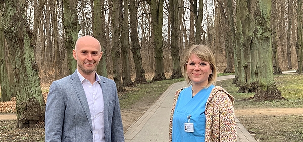
Rostock - Amyotrophic lateral sclerosis (ALS) is an incurable, neurodegenerative disease that leads to increasing paralysis of all muscles and death within 2-5 years. Due to the rapid progression of the disease and the rapidly developing immobility of the patients, it is challenging to provide them with the best possible care. The ALS Center at Rostock University Medicine is a central contact point for those affected in the region. With the support of the European Social Fund and the state government, a project is now being implemented that aims to provide interdisciplinary, networked care for all ALS sufferers in Mecklenburg-Western Pomerania (MV). In the future, patients and their families should be optimally cared for and regional and national resources should be consistently improved and utilized. In MV, up to 200 people are currently suffering from the incurable disease.
“The ALS patients and their relatives, some of whom are severely disabled, sometimes have great difficulty accessing specialized facilities in our country. This has a negative impact on the diagnosis and treatment of the disease and thus on the quality of life of patients and their families,” says Prof. Dr. Dr. Andreas Hermann, the Rostock study director. “We want to use new diagnostic and therapeutic options that are developing rapidly in the ALS field for patients as quickly as possible. The care structure in the country represents one of the biggest challenges. The offers must not be limited to a few patients in whose vicinity special consultations take place.
Starting from the ALS Center, the new case manager Sophie Fischer will set up an interdisciplinary, supra-regional care network in Mecklenburg-Western Pomerania as part of the funded project. She is the central contact person for all patients, doctors and therapists and will coordinate individual diagnostics and therapy as needed in accordance with the newly developed treatment paths. Sophie Fischer describes what cooperation in the country should look like in the future: “We are looking for practicing neurologists and general practitioners as well as neurological and internal medicine clinics who are willing to work with us to improve the care of ALS patients. We want to network with each other, develop a care concept together, increase the knowledge of patients, doctors and therapists and develop treatment standards.”
Also taking part is the MV regional association of the German Society for Muscular Diseases, the largest German self-help organization for neuromuscular diseases. Its honorary chairman Helmut Mädel is pleased that the development of the care network for ALS sufferers is being supported as a project. He hopes that there will also be a perspective afterwards: “The disease ALS does not end in two years. The biggest challenge will therefore be to be able to maintain the network on an independent financial basis after the two-year funding expires. It would also be desirable to expand the project to all neuromuscular diseases.”
There has been a specialized, academic ALS center in Rostock since 2008, which is known nationally and internationally. Rostock is also one of the eight founding members of the German Motor Neuron Network (MND-NET). There, proven ALS experts offer all aspects of state-of-the-art medicine, from initial diagnosis to diagnostic review and therapy. There are also disease-specific ALS self-help groups, family programs and clinical studies.
Contact:
Sophie Fischer
Neurological Polyclinic at the University of Rostock
Sophie.Fischer{bei}med.uni-rostock.de
(0381) 494 149542
There is a DGM contact group for patients with ALS (and other motor neuron diseases) and their relatives/carers. The meetings usually take place on Fridays 3 to 4 times a year. These meetings serve to exchange experiences with each other regarding this rare and fateful illness and also to provide socio-medical advice to patients and their relatives. As a rule, there is always an additional speaker. Many speakers from various specialist areas have already contributed their knowledge, experiences and recommendations to this round.
Details about this appointment can be found at:
Susanne Ulrich; E-mail: klaus-ulrich{bei}gmx.de; Tel.: 0381-710045
Katrin Körner; E-mail: katringast{bei}web.de; Tel: 0172 1605601
Sylvia Röhring; E-mail: sylvia.roehring{bei}web.de; Tel.: 0173 8810591
I look forward to our next meeting and will stay until then
With kind regards!
Sylvia Röhring
_____________
Dear members, dear fellow campaigners,
We would like to take this opportunity to send you our warmest New Year's greetings. We hope that you have had a good start to the new year and wish you all the best, many happy hours together with your loved ones and the strength to cope with everyday life.
We have planned joint meetings again for the new year and hope to be able to organise them regularly.
The dates for our contact group meetings are expected to be 15.03., 14.06., 20.09. and 15.11.2024. We have considered various topics for these meetings, including a presentation of aids by a medical supply store, information on the care degree and changes in care law.
We welcome ideas and requests for other topics and will endeavour to implement them.
The relatives' meetings are planned for 26.04. and 30.08.2024. All meetings will take place as usual at 5.00 pm in the St. Franziskus nursing home at Rudolf-Tarnow-Straße 12 in Rostock.
We are also planning to organise an ALS day. However, various arrangements still need to be made before this can take place.
We hope to see you again soon and send you
best regards
Susanne Ulrich, Katrin Körner and Sylvia Röhring
DGM: http://www.dgm.org/
DGN: https://dgn.org/leitlinie/motoneuronerkrankungen
Deutsches ALS Portal: http://www.als-site.de
ALS Charité: http://www.als-charite.de/
Deutsches Netzwerk für Motoneuronerkrankungen: http://www.mnd-als.de
Universitätszentrum für seltene Erkrankungen Rostock: https://selten.med.uni-rostock.de/
Augensteuerungs-Computer: http://www.interactive-minds.com/
Eine Patientin stellt sich vor: http://www.angelikahauffe.de/
"Initiative Therapieforschung ALS e.V." Dirk Peters, Dr. Hellmuth Stamm, Dr. Antje Tönnis (Vorstand) Gertrudenstr 31, 42105 Wuppertal (Deutschland) http://www.stop-als.de/

